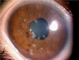
Feels like something is in the eye: Causes and treatment
What To Know About Flashes Of Light In The Corner Of The Eye
Seeing flashing lights in the corner of one or both eyes can occur with migraine or result from trauma, detached retina, or other problems. The flashes of light may vary in shape, color, frequency, and duration.
Flashes of light in the corner of the eye could be due to changes in the eye's structure, which becomes more common with age.
Some conditions, such as migraine auras, may also cause flashes of light in the eyes.
This article will examine the causes of flashes of light in the corner of the eye. It will also cover when a person should get advice from a doctor.
Flashes of light in the corner of the eye can result from an eye condition or injury. Photopsia is the medical name for these flashes, and this phenomenon usually occurs when there are changes inside the eye.
The retina is a thin layer of tissue that receives light at the back of the eye. It processes the light from the lens to send impulses through the optic nerve to the brain.
The vitreous body is a gel between the retina and lens that protects the retina and maintains the eye's structure.
According to the American Academy of Ophthalmology, most flashes occur when the vitreous body changes shape and pulls on the retina.
Occasional flashes are usually harmless and may happen more with aging. However, visual disturbances can also result from eye trauma, such as a blow to the eye or rubbing the eye too hard, or a medical condition.
Seeing flashes of light is not usually a cause for concern. However, if this occurs regularly, a person should contact a doctor.
Sometimes, flashes of light in the eye could signal a severe problem. They may also appear alongside floaters, which are tiny dots or lines that may appear in a person's vision.
The combination of sudden, repeated flashes with other visual disturbances could indicate vitreous detachment or a more severe condition.
Some eye-related causes of flashes in the corner of the eye can include:
Vitreous body or retinal damageChanges in the shape or position of the vitreous body are common and become more likely with age. A vitreous detachment can cause these flashes with floaters.
Vitreous detachment is a condition wherein the vitreous body breaks away from the retina. There are currently no treatments for vitreous detachment associated with aging, and people tend to adapt to the flashes and floaters eventually.
Vitreous detachment is not usually serious. However, it could have severe consequences, such as a hole or tear in the retina, for some people.
Tearing the retina can cause retinal detachment or bleeding in the eye. The symptoms can also include blurred or darkened vision.
Cryotherapy and laser therapy are common and effective treatments for retinal tears. For some people, however, the tear causes no symptoms and requires no treatment.
TraumaEye trauma can also cause flashes in the corner of the eye. Trauma can put pressure on the retina, causing flashes.
Symptoms of eye trauma might disappear immediately and require no treatment. However, a person should contact a doctor immediately if they experience any of the following symptoms:
A person should also avoid touching or scratching the eye.
Cytomegalovirus retinitisCytomegalovirus retinitis is a virus that affects the retinas. It can cause floaters with blurred vision that may lead to vision loss in one eye.
Without treatment, the symptoms of cytomegalovirus retinitis can spread to both eyes. The virus can also cause permanent retinal damage, resulting in blindness.
Treatments for cytomegalovirus retinitis include laser eye surgery and antiviral medications, such as ganciclovir (Cytovene).
Several other health conditions can cause flashes in the corner of the eye, such as:
MigrainePeople with migraine commonly experience auras. An aura is a collection of sensory disturbances that indicate the start of a migraine episode. These disturbances may include:
Combinations of medications that reduce the symptoms and prevent future episodes are available to treat migraine.
Occipital epilepsyOccipital epilepsy is a rare condition that affects some young children and teenagers with epilepsy. It may cause seizures that affect vision, leading to the person seeing flashing lights and multicolored spots.
Most young children and teenagers stop having these seizures as they age.
Doctors may treat occipital epilepsy using antiepileptic drugs to prevent seizures.
Stickler syndromeStickler syndrome is a rare genetic condition that can cause problems with the eyes, hearing, and joints. Stickler syndrome also commonly causes distinct facial features, such as a small chin and cleft palate.
Stickler syndrome can also cause eye abnormalities that increase the risk of developing retinal detachment, leading to flashes and floaters.
There is currently no cure for Stickler syndrome, and treatment depends on the specific symptoms a person experiences. For example, if a person has a detached retina, doctors may recommend laser surgery, freezing treatment, or other surgery types.
Diagnosis for flashing lights in the eyes will include an eye examination. A doctor will ask the person about their symptoms and any possible causes, such as a recent blow to the eye.
They will visually inspect the eye for any signs of injury, and they will also look out for distinctive features of someone with Stickler syndrome, such as a cleft palate.
An eye examination may include scleral depression, which involves applying gentle pressure to the eye. It may also involve using a specific lens for inspecting the retina.
A doctor may also use a dilated eye exam to check for cytomegalovirus retinitis. This involves using eye drops to dilate the eyes for inspection.
Flashes are an uncommon symptom of anxiety. Symptoms of anxiety typically include:
Some people report anxiety causing vision problems that include seeing stars or shimmers. However, there has been little research into visual disturbances as a symptom of anxiety.
Flashes in the corner of the eye can have many causes. The flashing mostly results from changes in the eye's structure, which becomes more likely with age. Age-related eye changes are usually harmless.
However, some causes of seeing flashes in the eyes could be severe. For example, retinal tears can cause bleeding or persistent vision problems. Some conditions, such as migraine auras, can also cause flashes in the eyes.
Anyone experiencing continuous flashing in the eyes or flashing alongside other visual disturbances should contact a doctor.
Evolution Of The Eye:
When evolution skeptics want to attack Darwin's theory, they often point to the human eye. How could something so complex, they argue, have developed through random mutations and natural selection, even over millions of years?
If evolution occurs through gradations, the critics say, how could it have created the separate parts of the eye -- the lens, the retina, the pupil, and so forth -- since none of these structures by themselves would make vision possible? In other words, what good is five percent of an eye?
Darwin acknowledged from the start that the eye would be a difficult case for his new theory to explain. Difficult, but not impossible. Scientists have come up with scenarios through which the first eye-like structure, a light-sensitive pigmented spot on the skin, could have gone through changes and complexities to form the human eye, with its many parts and astounding abilities.
Through natural selection, different types of eyes have emerged in evolutionary history -- and the human eye isn't even the best one, from some standpoints. Because blood vessels run across the surface of the retina instead of beneath it, it's easy for the vessels to proliferate or leak and impair vision. So, the evolution theorists say, the anti-evolution argument that life was created by an "intelligent designer" doesn't hold water: If God or some other omnipotent force was responsible for the human eye, it was something of a botched design.
Biologists use the range of less complex light sensitive structures that exist in living species today to hypothesize the various evolutionary stages eyes may have gone through.
Here's how some scientists think some eyes may have evolved: The simple light-sensitive spot on the skin of some ancestral creature gave it some tiny survival advantage, perhaps allowing it to evade a predator. Random changes then created a depression in the light-sensitive patch, a deepening pit that made "vision" a little sharper. At the same time, the pit's opening gradually narrowed, so light entered through a small aperture, like a pinhole camera.
Every change had to confer a survival advantage, no matter how slight. Eventually, the light-sensitive spot evolved into a retina, the layer of cells and pigment at the back of the human eye. Over time a lens formed at the front of the eye. It could have arisen as a double-layered transparent tissue containing increasing amounts of liquid that gave it the convex curvature of the human eye.
In fact, eyes corresponding to every stage in this sequence have been found in existing living species. The existence of this range of less complex light-sensitive structures supports scientists' hypotheses about how complex eyes like ours could evolve. The first animals with anything resembling an eye lived about 550 million years ago. And, according to one scientist's calculations, only 364,000 years would have been needed for a camera-like eye to evolve from a light-sensitive patch.
The Nocturnal Eye
 The Nocturnal Eye What appears as pitch black to a human may be dim light to a nocturnal animal. The reason lies in the structure of the eye itself.
Pupils
Nocturnal animals tend to have proportionally bigger eyes than humans do. They also tend to have pupils that open more widely in low light. So, at the outset, nocturnal eyes gather more light than human eyes do.Rods and cones
After the light passes through the pupil, it is focused by the lens onto the retina, which is connected to the brain by the optic nerve. The retina is an extremely complex structure. It's made up of at least 10 distinguishable layers, and is packed with more sensory nerve cells than anywhere else in the body.The retina is home to two different kinds of light receptor cells—rods and cones. (Both are named after their relative shapes.) Cones work in bright light and register detail, while rods work in low light, detecting motion and basic visual information. It is the rods that become highly specialized in nocturnal animals. In fact, many bats, nocturnal snakes and lizards have no cones at all, while other nocturnal animals have just a few.
Tapetum
Many nocturnal eyes are equipped with a feature designed to amplify the amount of light that reaches the retina. Called a tapetum, this mirror-like membrane reflects light that has already passed through the retina back through the retina a second time, giving the light another chance to strike the light-sensitive rods. Whatever light is not absorbed on this return trip passes out of the eye the same way it came in—through the pupil. The presence of the tapetum can be observed at night when a pair of glowing eyes reflects back a flashlight or some other light source. (Interestingly, different animals have different color tapeta, a fact that can aid in nighttime animal identification.)Circular vs. Slit pupils
One consequence of having extremely light sensitive eyes, is that they must be adequately protected during the day. Some animals accomplish this with a retractable eye flap. Others rely on their pupils.The circular pupil, because of the way the muscle bunches as it contracts, is the least efficient at closing rapidly and completely. A slit pupil, with two sides that can close like a sliding door, is far better at this task, which is why so many nocturnal eyes have slit pupils. These apertures can be vertical, horizontal, or diagonal.
Night VisionZoology After DarkResourcesGuideTranscriptNight Creatures Home
Editor's PicksPrevious SitesJoin Us/E-mailTV/Web ScheduleAbout NOVATeachersSite MapShopJobsSearchTo printPBS OnlineNOVA OnlineWGBH©Updated November 2000


Comments
Post a Comment